{{ isErrorSetToBasket === false ? 'Товар добавлен вкорзину' : 'Не удалось добавить товар в корзину'}}
Перейти в корзину
{{Object.keys(error)[0]}}:
{{Object.values(error)[0]}}
Цена По запросу
Количество
Вы уже добавили максимально доступное на складе кол-во товара
Достигнуто максимально доступное кол-во
Под заказ
{{!!storageProduct ? 'На складе' : 'Под заказ'}}
Ожидается поставка
Description
General description
Neurofilament heavy polypeptide (UniProt: P12036; also known as NF-H, 200 kDa neurofilament protein, Neurofilament triplet H protein) is encoded by the NEFH (also known as KIAA0845, NFH) gene (Gene ID: 4744) in human. Neurofilaments (~ 10 nm in diameter) are intermediate filaments that serve as major elements of the cytoskeleton supporting the axon cytoplasm. They are the most abundant fibrillar components of the axon, being on average 3-10 times more frequent than axonal microtubules. They are built from three intertwined protofibrils, which are themselves composed of two tetrameric protofilament complexes of monomeric proteins. The neurofilament triplet proteins (68/70, 160, and 200 kDa) occur in both the central and peripheral nervous system and are usually neuron specific. The 68/70 kDa NF-L protein can self-assemble into a filamentous structure, however the 160 kDa NF-M and 200 kDa NF-H proteins require the presence of the 68/70 kDa NF-L protein to co-assemble. Antibodies to NF-H are useful for identifying neuronal cells and their processes in tissue sections and in tissue culture. NF-H antibodies are helpful in the identification of neurofilament accumulations seen in many neurological diseases, such as Amyotrophic lateral sclerosis (ALS) and Alzheimer′s disease. The purified immunogen used to generate this antibody was the very heavily phosphorylated form of NF-H and this antibody binds only this heavily phosphorylated form. Similar, but far fewer phosphorylation sites are also found in NF-M. Hence, this antibody displays some reactivity with NF-M. Enzymatic phosphorylation of porcine and bovine neurofilaments is shown to result in loss of binding.
Specificity
Clone NP1 detects heavily phosphorylated form of Neurofilament H. Displays weaker reactivity with phosphorylated Neurfilament -M.
Immunogen
Native Neurofilament H purified from pig spinal cord.
Application
Anti-Neurofilament NF-H, phosphorylated, clone NP1, Cat. No. MAB1592-C, is a highly specific mouse monoclonal antibody that targets Neurofilament heavy polypeptide and has been tested for use in ELISA, Immunohistochemistry (Paraffin), and Western Blotting.
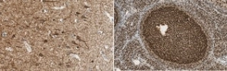
Immunohistochemistry Analysis: A 1:50 dilution from a representative lot detected Neurofilament NF-H in human ovary tissue.
Immunohistochemistry Analysis: A representative lot detected Neurofilament NF-H in Immunohistochemistry applications (Benson, D.L., et. al. (1996). J Neurocytol. 25(3):181-96; Boylan, K., et. al. (2009). J Neurochem. 111(5):1182-91).
Western Blotting Analysis: A representative lot detected Neurofilament NF-H in Western Blotting applications (Benson, D.L., et. al. (1996). J Neurocytol. 25(3):181-96; Boylan, K., et. al. (2009). J Neurochem. 111(5):1182-91).
ELISA Analysis: A representative lot detected Neurofilament NF-H in ELISA applications (Boylan, K., et. al. (2009). J Neurochem. 111(5):1182-91; Toedebusch, C.M., et. al. (2017). J Vet Intem Med. 31(2):513-520; Mashita, T., et. al. (2015). J Vet Med Sci. 77(4):433-8).
Research Category
Neuroscience
Target description
112.48 kDa calculated.
Physical form
Format: Purified
Protein G purified
Purified mouse monoclonal antibody IgG1 in PBS with 5 mM sodium azide and 50% glycerol.
Storage and Stability
Stable for 1 year at -20°C from date of receipt.
Handling Recommendations: Upon receipt and prior to removing the cap, centrifuge the vial and gently mix the solution. Aliquot into microcentrifuge tubes and store at -20°C. Avoid repeated freeze/thaw cycles, which may damage IgG and affect product performance.
Disclaimer
Unless otherwise stated in our catalog or other company documentation accompanying the product(s), our products are intended for research use only and are not to be used for any other purpose, which includes but is not limited to, unauthorized commercial uses, in vitro diagnostic uses, ex vivo or in vivo therapeutic uses or any type of consumption or application to humans or animals.
Quality
Evaluated by Immunohistochemistry in human cerebral cortex tissue.
Immunohistochemistry Analysis: A 1:50 dilution of this antibody detected Neurofilament NF-H in human cerebral cortex tissue.
Other Notes
Concentration: Please refer to lot specific datasheet.
- Related Categories Alphabetical Index, Antibodies, NE-NI, Primary Antibodies clone